| Спинномозговой нерв | |
|---|---|
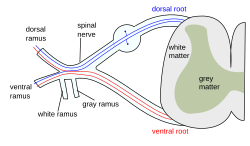
 Формирование спинномозгового нерва из заднего и переднего корешков | |
| Подробности | |
| Идентификаторы | |
| латинский | спинной нерв |
| MeSH | D013127 |
| TA98 | А14.2.00.027 А14.2.02.001 |
| TA2 | 6143 , 6362 |
| FMA | 5858 |
| Анатомические термины нейроанатомии | |
Спинной нерв представляет собой смешанный нерв , который несет двигатель, сенсорный и вегетативные сигналы между спинным мозгом и телом. В теле человека 31 пара спинномозговых нервов, по одной с каждой стороны позвоночника . Они сгруппированы в соответствующие шейные , грудные , поясничные , крестцовые и копчиковые области позвоночника. [1] Есть восемь пар шейных нервов , двенадцать пар грудных нервов , пять пар поясничных нервов , пять пар крестцовых нервов., и одна пара копчиковых нервов . Спинномозговые нервы являются частью периферической нервной системы .
Структура [ править ]
Каждый спинномозговой нерв представляет собой смешанный нерв, образованный комбинацией нервных волокон его дорсального и вентрального корешков . Спинной корешок является афферентным сенсорным корнем и передает сенсорную информацию в мозг. Брюшной корешок является эфферентным моторным корнем и несет моторную информацию от мозга. Спинной нерв выходит из позвоночника через отверстие ( межпозвонковое отверстие ) между соседними позвонками. Это верно для всех спинномозговых нервов, за исключением первой пары спинномозговых нервов (C1), которая проходит между затылочной костью и атлантом.(первый позвонок). Таким образом, шейные нервы нумеруются нижним позвонком, за исключением спинномозгового нерва C8, который находится ниже позвонка C7 и выше позвонка T1. Затем грудной, поясничный и крестцовый нервы нумеруются по указанному выше позвонку. В случае поясничного позвонка S1 (также известного как L6) или сакрализованного позвонка L5, нервы обычно по-прежнему считаются до L5, а следующий нерв - S1.
1. Соматический эфферент .
2. Соматический афферент .
3,4,5. Симпатический эфферент .
6,7. Вегетативный афферент .
Вне позвоночника нерв делится на ветви. Спинная ветвь содержит нервы , которые служат задние части ствола , несущих висцерального двигатель, двигатель, соматический и соматический сенсорную информацию к и от кожи и мышц спины ( расположенный выше или позади оси мышцы ). Вентральная ветвь содержит нервы , которые служат оставшиеся передние части туловища и верхних и нижних конечности ( расположенные спереди от вертикальной оси тела мышц ) , несущих висцерального двигатель, соматической двигатель, и сенсорной информации к и от вентролатеральной поверхности тела, структуры в стенке корпуса, и конечности. В менингеальные ветви (рецидивирующий менингеальные или sinuvertebral нервов) branch from the spinal nerve and re-enter the intervertebral foramen to serve the ligaments, dura, blood vessels, intervertebral discs, facet joints, and periosteum of the vertebrae. The rami communicantes contain autonomic nerves that serve visceral functions carrying visceral motor and sensory information to and from the visceral organs.
Some anterior rami merge with adjacent anterior rami to form a nerve plexus, a network of interconnecting nerves. Nerves emerging from a plexus contain fibers from various spinal nerves, which are now carried together to some target location. Major plexuses include the cervical, brachial, lumbar, and sacral plexuses.
Регионарные нервы [ править ]
Шейные нервы [ править ]
Шейные нервы - это спинномозговые нервы от шейных позвонков в шейном сегменте спинного мозга. Хотя есть семь шейных позвонков (С1-С7), существует восемь шейных нервов C1 - C8 . С1 – С7 выходят над соответствующими позвонками, а С8 - под позвонком С7. Повсюду в позвоночнике нерв выходит под позвонком с тем же названием.
Заднее распределение включает подзатылочный нерв (C1), большой затылочный нерв (C2) и третий затылочный нерв (C3). Переднее распределение включает шейное сплетение (C1-C4) и плечевое сплетение (C5-T1).
Шейные нервы иннервируют грудинно - подъязычную , sternothyroid и omohyoid мышцы .
Петля нервов, называемая ansa cervicalis, является частью шейного сплетения.
Грудные нервы [ править ]
The thoracic nerves are the twelve spinal nerves emerging from the thoracic vertebrae. Each thoracic nerve T1 -T12 originates from below each corresponding thoracic vertebra. Branches also exit the spine and go directly to the paravertebral ganglia of the autonomic nervous system where they are involved in the functions of organs and glands in the head, neck, thorax and abdomen.
Anterior divisions: The intercostal nerves come from thoracic nerves T1-T11, and run between the ribs. At T2 and T3, further branches form the intercostobrachial nerve. The subcostal nerve comes from nerve T12, and runs below the twelfth rib.
Задние отделы: медиальные ветви (ramus medialis) задних ветвей шести верхних грудных нервов проходят между semispinalis dorsi и multifidus , которые они снабжают; Затем они прокалывают ромбовидные и трапециевидные мышцы и достигают кожи по бокам остистых отростков. Эта чувствительная ветвь называется медиальной кожной ветвью.
Медиальные ветви шести нижних мышц распространяются в основном на multifidus и longissimus dorsi , иногда они отдают нити на кожу около средней линии. Эта чувствительная ветвь называется задней кожной ветвью.
Поясничные нервы [ править ]
The lumbar nerves are the five spinal nerves emerging from the lumbar vertebrae. They are divided into posterior and anterior divisions.
Posterior divisions: The medial branches of the posterior divisions of the lumbar nerves run close to the articular processes of the vertebrae and end in the multifidus muscle.
The laterals supply the erector spinae muscles.
The upper three give off cutaneous nerves which pierce the aponeurosis of the latissimus dorsi at the lateral border of the erector spinae muscles, and descend across the posterior part of the iliac crest to the skin of the buttock, some of their twigs running as far as the level of the greater trochanter.
Anterior divisions: The anterior divisions of the lumbar nerves (rami anteriores) increase in size from above downward. They are joined, near their origins, by gray rami communicantes from the lumbar ganglia of the sympathetic trunk. These rami consist of long, slender branches which accompany the lumbar arteries around the sides of the vertebral bodies, beneath the psoas major. Their arrangement is somewhat irregular: one ganglion may give rami to two lumbar nerves, or one lumbar nerve may receive rami from two ganglia.
The first and second, and sometimes the third and fourth lumbar nerves are each connected with the lumbar part of the sympathetic trunk by a white ramus communicans.
The nerves pass obliquely outward behind the psoas major, or between its fasciculi, distributing filaments to it and the quadratus lumborum.
The first three and the greater part of the fourth are connected together in this situation by anastomotic loops, and form the lumbar plexus.
The smaller part of the fourth joins with the fifth to form the lumbosacral trunk, which assists in the formation of the sacral plexus. The fourth nerve is named the furcal nerve, from the fact that it is subdivided between the two plexuses.
Sacral nerves[edit]
The sacral nerves are the five pairs of spinal nerves which exit the sacrum at the lower end of the vertebral column. The roots of these nerves begin inside the vertebral column at the level of the L1 vertebra, where the cauda equina begins, and then descend into the sacrum.[2][3]
There are five paired sacral nerves, half of them arising through the sacrum on the left side and the other half on the right side. Each nerve emerges in two divisions: one division through the anterior sacral foramina and the other division through the posterior sacral foramina.[4]
The nerves divide into branches and the branches from different nerves join with one another, some of them also joining with lumbar or coccygeal nerve branches. These anastomoses of nerves form the sacral plexus and the lumbosacral plexus. The branches of these plexus give rise to nerves that supply much of the hip, thigh, leg and foot.[5][6]
The sacral nerves have both afferent and efferent fibers, thus they are responsible for part of the sensory perception and the movements of the lower extremities of the human body. From the S2, S3 and S4 arise the pudendal nerve and parasympathetic fibers whose electrical potential supply the descending colon and rectum, urinary bladder and genital organs. These pathways have both afferent and efferent fibers and, this way, they are responsible for conduction of sensory information from these pelvic organs to the central nervous system (CNS) and motor impulses from the CNS to the pelvis that control the movements of these pelvic organs.[7]
Coccygeal nerve[edit]
The coccygeal nerve is the 31st pair of spinal nerves. It arises from the conus medullaris, and its anterior root helps form the coccygeal plexus. It does not divide into a medial and lateral branch. It is distributed to the skin over the back of the coccyx.
Function[edit]
| Level | Motor function |
|---|---|
| C1–C6 | Neck flexors |
| C1–T1 | Neck extensors |
| C3, C4, C5 | Supply diaphragm (mostly C4) |
| C5, C6 | Move shoulder, raise arm (deltoid); flex elbow (biceps) |
| C6 | externally rotate (supinate) the arm |
| C6, C7 | Extend elbow and wrist (triceps and wrist extensors); pronate wrist |
| C7, C8 | Flex wrist; supply small muscles of the hand |
| T1–T6 | Intercostals and trunk above the waist |
| T7–L1 | Abdominal muscles |
| L1–L4 | Flex hip joint |
| L2, L3, L4 | Adduct thigh; Extend leg at the knee (quadriceps femoris) |
| L4, L5, S1 | abduct thigh; Flex leg at the knee (hamstrings); Dorsiflex foot (tibialis anterior); Extend toes |
| L5, S1, S2 | Extend leg at the hip (gluteus maximus); flex foot and flex toes |
Clinical significance[edit]
The muscles that one particular spinal root supplies are that nerve's myotome, and the dermatomes are the areas of sensory innervation on the skin for each spinal nerve. Lesions of one or more nerve roots result in typical patterns of neurologic defects (muscle weakness, abnormal sensation, changes in reflexes) that allow localization of the responsible lesion.
There are several procedures used in sacral nerve stimulation for the treatment of various related disorders.
Sciatica is generally caused by the compression of lumbar nerves L4, or L5 or sacral nerves S1, S2, or S3, or by compression of the sciatic nerve itself
Additional Images[edit]
A portion of the spinal cord, showing its right lateral surface. The dura is opened and arranged to show the nerve roots.
Distribution of the cutaneous nerves. Ventral aspect.
Distribution of the cutaneous nerves. Dorsal aspect.
The spinal cord with dura cut open, showing the exits of the spinal nerves.
The spinal cord showing how the anterior and posterior roots join in the spinal nerves.
A longer view of the spinal cord.
Projections of the spinal cord into the nerves (red motor, blue sensory).
Projections of the spinal cord into the nerves (red motor, blue sensory).
Schematic diagram of cervical plexus.
- Dissection images
Cerebrum. Inferior view. Deep dissection.
Cerebrum. Inferior view. Deep dissection.
Spinal nerves. Spinal cord and vertebral canal. Deep dissection.
See also[edit]
- Cranial nerves
References[edit]
- ^ "Spinal Nerves". National Library of Medicine. Retrieved 12 April 2014.
- ^ 1. Anatomy, descriptive and surgical: Gray's anatomy. Gray, Henry. Philadelphia : Courage Books/Running Press, 1974
- ^ 2. Clinically Oriented Anatomy. Moore, Keith L. Philadelphia : Wolters Kluwer Health/Lippincott Williams & Wilkins, 2010 (6th ed)
- ^ 1. Anatomy, descriptive and surgical: Gray's anatomy. Gray, Henry. Philadelphia : Courage Books/Running Press, 1974
- ^ 1. Anatomy, descriptive and surgical: Gray's anatomy. Gray, Henry. Philadelphia : Courage Books/Running Press, 1974
- ^ 3. Human Neuroanatomy. Carpenter, Malcolm B. Baltimore : Williams & Wilkins Co., 1976 (7th ed)
- ^ 3. Human Neuroanatomy. Carpenter, Malcolm B. Baltimore : Williams & Wilkins Co., 1976 (7th ed)
- Blumenfeld H. 'Neuroanatomy Through Clinical Cases'. Sunderland, Mass: Sinauer Associates; 2002.
- Drake RL, Vogl W, Mitchell AWM. 'Gray's Anatomy for Students'. New York: Elsevier; 2005:69-70.
- Ropper AH, Samuels MA. 'Adams and Victor's Principles of Neurology'. Ninth Edition. New York: McGraw Hill; 2009.

