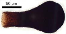
Chitinozoan
Chitinozoa (singular: chitinozoan, plural: chitinozoans) are a group of flask-shaped, organic walled marine microfossils produced by an as yet unknown organism.[1] Common from the Ordovician to Devonian periods (i.e. the mid-Paleozoic), the millimetre-scale organisms are abundant in almost all types of marine sediment across the globe.[2] This wide distribution, and their rapid pace of evolution, makes them valuable biostratigraphic markers.[3][4]
Their bizarre form has made classification and ecological reconstruction difficult. Since their discovery in 1931, suggestions of protist, plant, and fungal affinities have all been entertained. The organisms have been better understood as improvements in microscopy facilitated the study of their fine structure, and it has been suggested that they represent either the eggs or juvenile stage of a marine animal.[5] However, recent research has suggested that they represent the hard shell of a group of protists with uncertain affinities.[6]
Chitinozoan ecology is also open to speculation; some may have floated in the water column, where others may have attached themselves to other organisms. Most species were particular about their living conditions, and tend to be most common in specific paleoenvironments. Their abundance also varied with the seasons.
Chitinozoa range in length from around 50 to 2000 micrometres.[2] They appear dark to almost opaque when viewed under an optical microscope. Their anatomy is based around the broad chamber, a radially symmetrical region involving a central cavity encased by two layers of a chitin-like substance. The chamber narrows towards the main opening (the aperture), though a circular plug prevents direct contact between the central cavity and its surroundings. This plug may be called an operculum (if it lies at the tip of the aperture) or a prosome (if it lies deep within the narrowed region or neck). The rim of the aperture, known as the collarette, often has a distinctive form or texture.[3]
The base of the chitinozoan lies at the opposite end from the aperture. The base may involve various ornamentation derived from the internal layer. The edge of the base (basal margin) may extend into a sharp radial plate, the carina. Alternatively, it could send out large spines or branches, known as processes. In chitinozoans which attach to substrates or each other in large chains, the center of the base is augmented with apical structures which project down to assist attachment.[3]
External ornamentation is often preserved on the surface of the fossils, in the form of hairs, loops or protrusions, which are sometimes as large as the chamber itself. The range and complexity of ornament increased with time, against a backdrop of decreasing organism size. The earliest Ordovician species were large and smooth-walled;[7] by the mid-Ordovician a large and expanding variety of ornament, and of hollow appendages, was evident. While shorter appendages are generally solid, larger protrusions tend to be hollow, with some of the largest displaying a spongy internal structure.[8] However, even hollow appendages leave no mark on the inner wall of the organisms: this may suggest that they were secreted or attached from the outside.[8] There is some debate about the number of layers present in the organisms' walls: up to three layers have been reported, with the internal wall often ornamented; some specimens only appear to display one such wall layer. The multitude of walls may indeed reflect the construction of the organism, but could be a result of the preservational process.[8]


