Hip
In vertebrate anatomy, the hip, or coxa[1](pl.: coxae) in medical terminology, refers to either an anatomical region or a joint on the outer (lateral) side of the pelvis.
The hip region is located lateral and anterior to the gluteal region, inferior to the iliac crest, and lateral to the obturator foramen, with muscle tendons and soft tissues overlying the greater trochanter of the femur.[2] In adults, the three pelvic bones (ilium, ischium and pubis) have fused into one hip bone, which forms the superomedial/deep wall of the hip region.
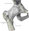
The hip joint, scientifically referred to as the acetabulofemoral joint (art. coxae), is the ball-and-socket joint between the pelvic acetabulum and the femoral head. Its primary function is to support the weight of the torso in both static (e.g. standing) and dynamic (e.g. walking or running) postures. The hip joints have very important roles in retaining balance, and for maintaining the pelvic inclination angle.
Pain of the hip may be the result of numerous causes, including nervous, osteoarthritic, infectious, traumatic, and genetic.
The hip joint, also known as a ball and socket joint, is formed by the acetabulum of the pelvis and the femoral head, which is the top portion of the thigh bone (femur). It allows for a wide range of movement and stability in the lower body.[3]
The proximal femur is largely covered by muscles and, as a consequence, the greater trochanter is often the only palpable bony structure in the hip region.[4]


